Echinococcus Granulosus, Hydatid Disease, Hydatid Cyst
Echinococcus Granulosus (Hydatid Disease)
What Sample is needed for Echinococcus Granulosus?
- A Haemagglutination test is done on the serum.
- Hydatid cyst fluid for the examination.
- The other specimens may be sputum, urine, liver, and spleen.
What are the Indications for Echinococcus Granulosus?
- This test is done to diagnose a parasitic disease caused by Echinococcus granulosus.
How will you describe the Biology of Echinococcus Granulosus?
- The genus Echinococcus contains three species:
- Echinococcus granulosus.
- Echinococcus multilocularis.
- Echinococcus vogeli.
- Humans are hosts in the larval stage or hydatid.
- The genus Echinococcus has the smallest tapeworm in the Taeniidae.
- The adult worm measures 0.6 cm or less in length.
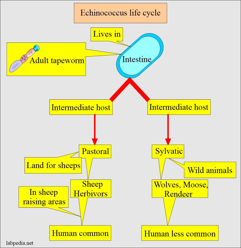
- This is found in areas where there are sheep (herbivorous).
- These are in close contact with the dogs and other canines.
- Other regions are where canines and humans have close contact.
How will you describe the Lifecycle of Echinococcus Granulosus?
- E. granulosus is a parasitic disease caused by a tapeworm of the genus Echinococcus.
- This is also called Hydatid cyst disease and Hydatid cyst.
What are the features of the adult worm?
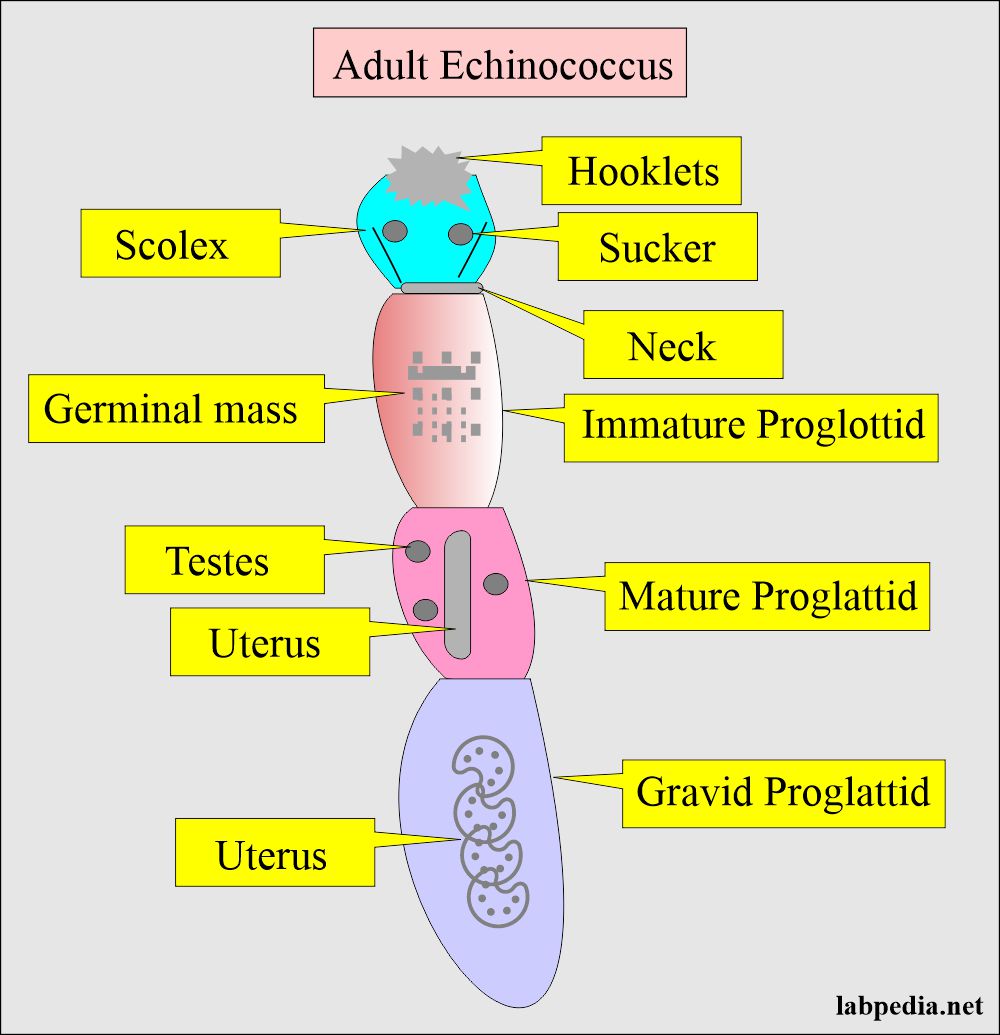
- It is 0.6 cm (3 to 8mm) or less in length.
- It consists of the following:
- Scolex.
- Neck.
- Proglottids are 3 in number (the segment containing sex organs) and are:
- One anterior-most is immature.
- One is middle and is mature.
- One is terminal and is gravid.
What are the hosts for the spread of Echinococcus Granulosis?
- Definitive host:
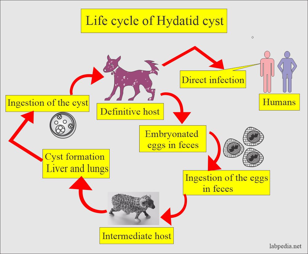
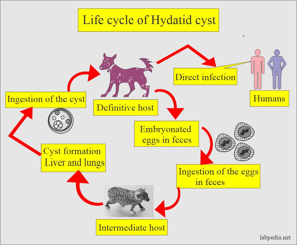
- E. granulosus requires two mammalian hosts (animal likess dogs, sheep, horses, goats, pigs, and cattle) to complete their life cycle.
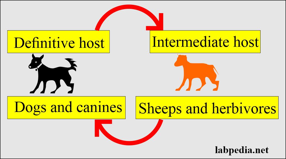
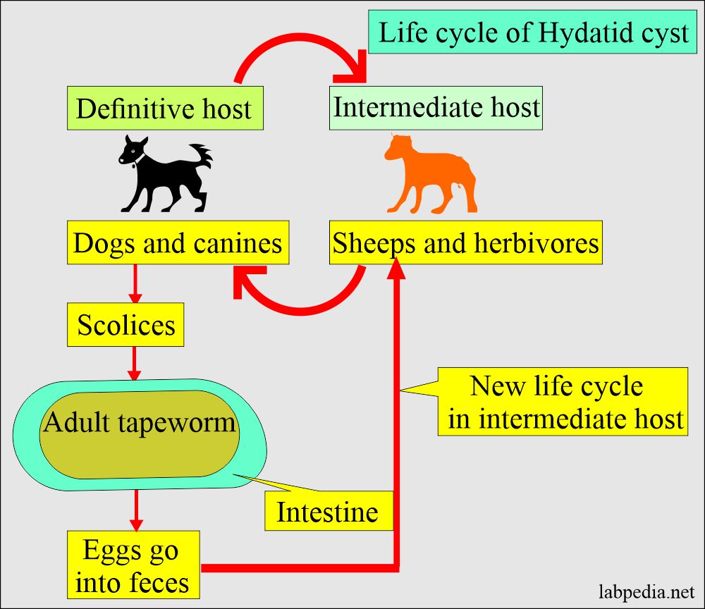
- It uses dogs or other canines as a definitive host.
- The dogs are the definitive host.
- Sheep and cattle are intermediate hosts.
- Infection is commonest in sheep and cattle-raising areas.
- Habitat is a small intestine.
- It uses dogs or other canines as a definitive host.
- Intermediate host:
- Herbivorous is an intermediate host that becomes infected by eating the contaminated herbage.
- Segments containing eggs (gravid proglottids) or free eggs are passed in the feces of the definitive host, a carnivore.
- The eggs are ingested by an intermediate host (maybe humans), in which the metacestode stage and protoscoleces develop.
- Then, the cycle is completed if a suitable carnivore eats the metacestode and protoscoleces.
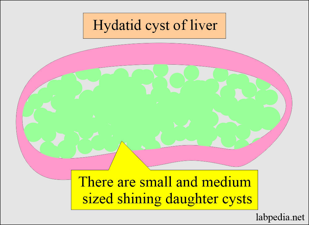
- Hydatid cyst (Hydatidosis) develops in various organs. The liver and lungs are the most common sites. Still, it can be seen on any other site; even these cysts are reported in the eye, brain, kidney, spleen, muscles, and bones.
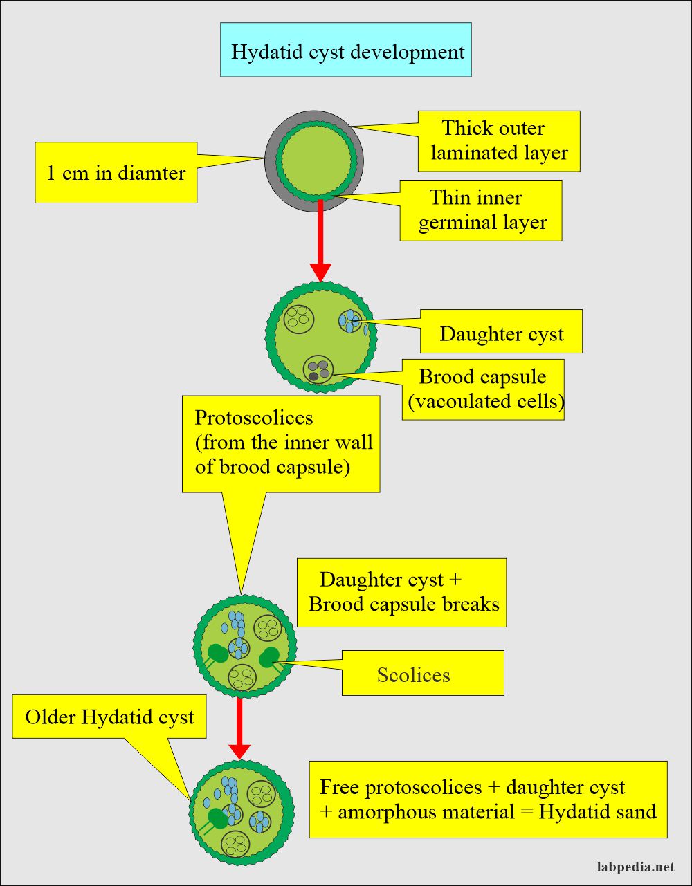
- Hydatid cyst in the early stages is out of the thick laminated membrane and lined by thin germinal epithelium; its size, in the beginning, is 1 cm in diameter.
- In the end, the older hydatid cyst contains free protoscolices, a daughter cyst, and an amorphous material, and all these are called hydatid sand.
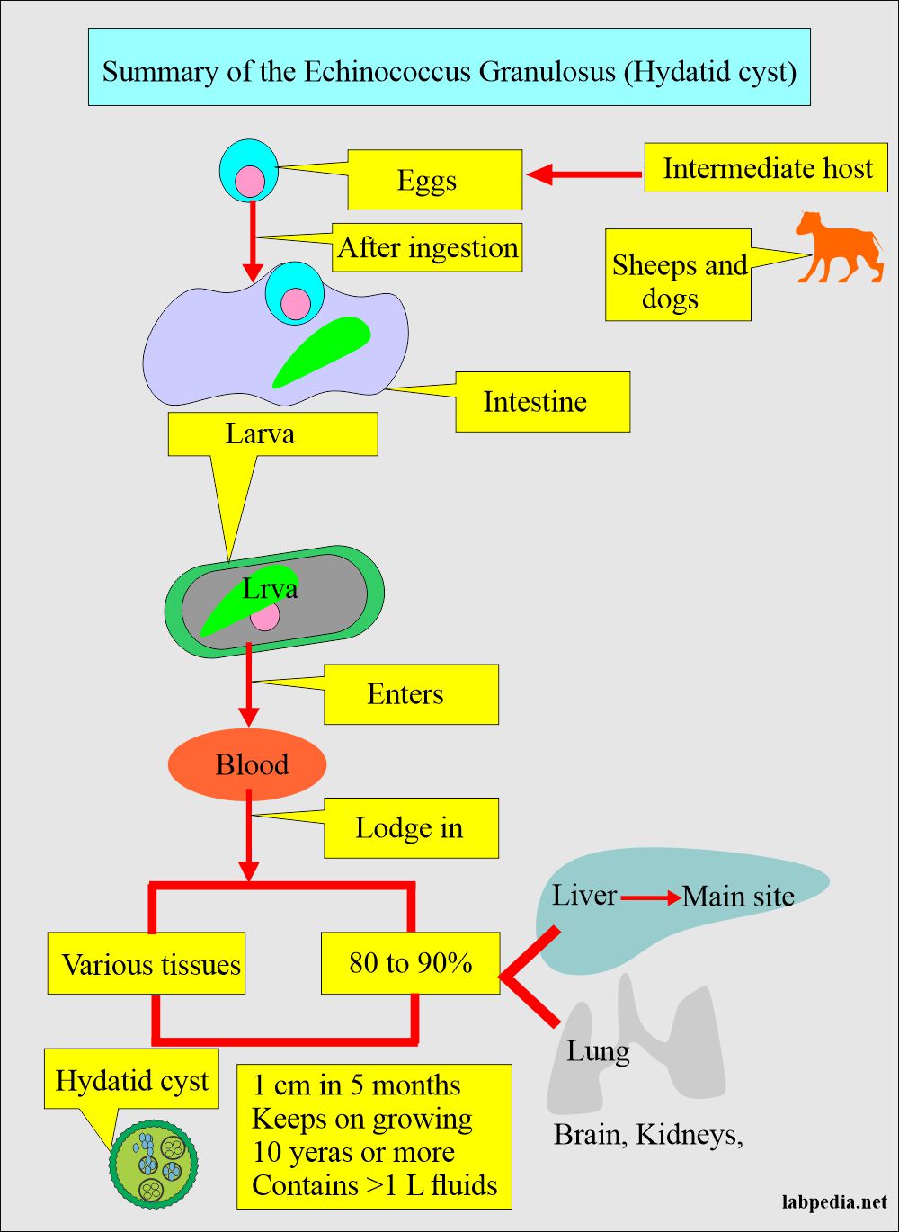
How will you describe the Summary of the life cycle of Echinococcus granulosus (Hydatid cyst)?
- Humans ingest eggs from sheep and dogs.
- These eggs will hatch in the intestine and form larvae.
- The larva penetrates the wall of the intestine and enters the blood circulation.
- The majority settle in the liver, and a few go to the brain, lungs, and kidneys.
- Rarely are these found in unexpected places like the eye, vagina, etc.
- These larvae form the hydatid cyst, which has fluid in the center. There is the formation of daughter hydatid cysts and protoscolices in the cysts.
- The size may reach 5 to 10 cm.
- This multilocular cyst is called Echinococcus multilocularis.
- 10% of the cysts cause symptoms and are difficult to treat.
- If the cyst fluid leaks out, it causes a fatal allergic reaction.
What is the Epidemiology of Echinococcus Granulosus?
- This disease is present throughout the world.
- Children are more likely to get an infection due to poor hygienic habits.
- Human infection occurs accidentally through the feces of infected dogs or other herbivores.
- These prevail in sheep-raising areas like New Zealand and Australia.
- This disease is quite common in the South American continent.
- It is seen in Alaska, Canada, and the nearby United States in North America.
What are the Signs and Symptoms of Echinococcus Granulosus?
- 10% of the cysts cause signs and symptoms.
- This depends upon the size and location of the hydatid cyst.
- The patients are asymptomatic; the incubation period may be in years when the cyst appears and is clinically diagnosed.
- There are nonspecific symptoms of:
- Nausea.
- Weight loss.
- Weakness.
- Other symptoms are due to the pressure of these cysts.
- In the case of the liver cyst:
- The patient may have abdominal pain.
- Nausea and vomiting.
- There may be obstructive jaundice.
- In the case of the lung:
- There will be a chronic cough.
- Chest pain and shortness of breath.
- Rupture of the cyst naturally or by taking the sample may lead to anaphylactic shock.
- Summary of signs and symptoms:
- Asymptomatic infection.
- Hepatomegaly.
- Cough.
- Pressure on different organs.
- Rupture of the cyst may cause allergic reactions like anaphylactic reactions.
How will you Diagnose Echinococcus Granulosus?
- A complete blood picture may show eosinophilia.
- Haemagglutination test with significant titer above 1:100.
- Indirect hemagglutination and ELISA are also satisfactory tests.
- Serological tests are more specific than the traditional Casoni test.
- Direct microscopic examination for ova and parasites.
- Microscopy of hydatid cyst fluid shows scolices, daughter cysts, brood capsules, or hydatid sand.
- X-rays may show cysts in asymptomatic patients. The cyst shows a sharp outline and may see fluid levels.
- Bone marrow biopsy.
- CT scan or ultrasonography helps find a hydatid cyst.
How will you treat the Echinococcus Granulosus?
- Surgical removal of the operable cyst. This is the best choice of treatment.
- In the case of inoperable cases, can give:
- Mebendazole.
- Albendazole.
- Praziquantel.
- The first drug, Mebendazole, was found to be effective.
- Albendazole is given for a longer time to kill the cyst. This was better absorbed and penetrated the cyst wall. Now, this drug is preferred to mebendazole.
- Albendazole is given as 10 mg/Kg body weight or 400 mg twice daily for 4 weeks.
- This cycle can be repeated at 2-week intervals. You can give such 12 cycles of Albendazole drugs.
- Response to therapy can be judged by ultrasound or MRI and may be repeated at intervals of 3 months.
- Percutaneous injection of hypertonic saline (solicidal agents) and aspiration is sometimes considered safe and effective therapy.
- Praziquantel is protoscilocidal drug. It has a very good result when given with Albendazole.
- Can inject saline, iodophor, and alcohol to ensure that the cystic contents are dead.
- After 30 minutes, it can be enucleated from the cyst.
How will you Prevent Echinococcus Granulosus?
- Improve the hygiene of people handling the dogs.
- Frequently treat the dogs in endemic areas.
- Improve the ways for the disposal of the dog’s feces.
- 6 weekly deworming of the dogs with praziquantel is very effective.
Questions and answers:
Question 1: What is the definitive host of the Echinococcus Granulosus?
Question 2: What are sites for the deposition of hydatid cysts?

