White blood cell:- Part 2 – Total Leukocytes Count Procedure, TLC Solution Preparation
White blood cell
Total leucocytes count procedure (TLC)
Sample
- EDTA blood is needed.
- Oxalate or citrated blood can also be used.
Purpose of the test (Indications) for Total leucocytes count
- To differentiate between acute and chronic infection.
- To follow the patient with chemotherapy.
- To find the effect of drugs.
Principle of white cell count (total leucocytes count)
Blood is diluted with a solution (acetic acid) that causes the lysis of RBCs. Acetic acid has no effect on the WBCs. Gentian violet is added to differentiate the WBCs.
Pathophysiology of Total leucocytes count TLC)
- White blood cells develop from the stem cells in the bone marrow.
- Stem cells differentiate into:
- Granulocytic series cells. The granulocytes got the name due to the presence of distinct granules in the cytoplasm of these cells.
- The life span of leucocytes varies from 13 to 20 days; after that, these cells are destroyed in the lymphatic system and may be excreted from the body in feces.
- Leucocytes fight infections and defend the body by a process called phagocytosis.
- WBC serves as an excellent indicator of the various disease process.
- Non-Granulocytic cells.
- The granulocytic series consists of:
- polymorphonuclear leucocytes (Neutrophils).
- Eosinophils.
- Basophils.
- Monocytes.
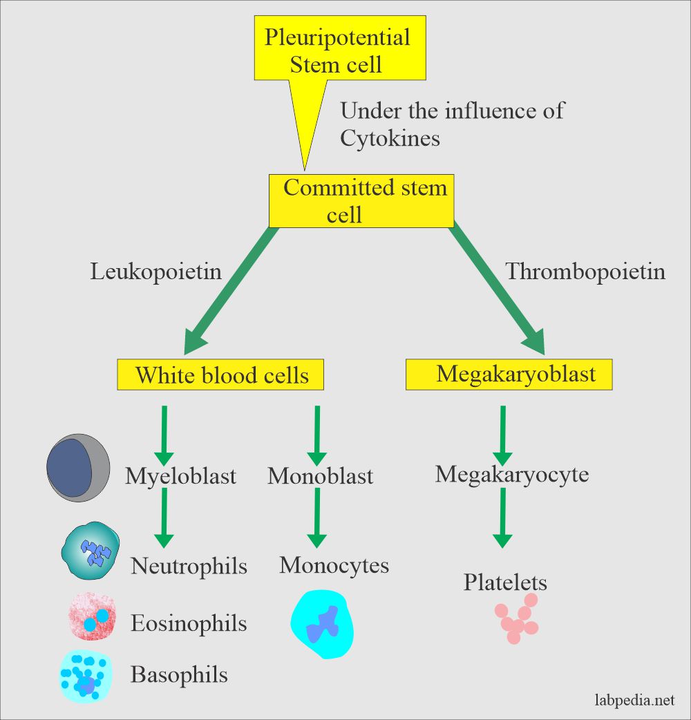
Development of white blood cells
- Granulocytic series cells. The granulocytes got the name due to the presence of distinct granules in the cytoplasm of these cells.
White blood cells are divided into:
- Phagocytic cells:
- Polys.
- Eosinophils.
- Basophils.
- Monocytes.
- Immunocytic cells:
- B- lymphocytes.
- T-lymphocytes are:
- T-helper cells (CD4+)
- T-cytotoxic cells (CD+)
- T-effector cells (CD4+)
Preparation of Total leucocytes count (TLC) solution:
- Principle of the TLC method:
- Blood is diluted with a fluid that causes the RBCs’ hemolysis, but WBCs remain intact, and then these are counted in the Neubauer chamber.
- Gentian violet lightly stains the leucocytes and allows those to be counted.
- Chemicals (Reagents) needed are:
- Glacial acetic acid = 2 ml.
- Gentian violet (1%) = 1 ml.
- Distal water = 97 ml.
- This is basically a 2% solution of acetic acid.
- Add 2 ml of glacial acid (2 + 1), 1 mL gentian violet (1 %), and add 97 ml of distle Water to make up to 100 ml solution.
- Gentian violet is added until the color is pale blue-violet.
- Total leucocyte count or WBC pipette:
- This pipette (also called Thoma pipette) long stem is divided into two parts:
- The long stem is marked with 0.5 and 1.0
- While the short arm after the bulb is marked 11.
- Its central portion is a bulb or a globular shape with one white bead in it.
- Rubber tubing is attached to suck the blood.
- Ultimately the dilution of the blood to the TLC fluid is 1:20.

WBC pipette
- This pipette (also called Thoma pipette) long stem is divided into two parts:
Total leucocytes count Procedure:
- Take the TLC pipette, which has a white bead inside.
- Total leucocytes count (WBC) pipette method:
- Fill the blood into the 0.5 marks and then add the TLC solution.
- Fill the pipette with the TLC solution to point 11.
- Remove the rubber tubing.
- Seal both ends or hold in between two fingers.
- OR can put this pipette on the mechanical device to shake it.
- Shake for 1 minute or preferably for 2 minutes.
- Shaking is important before filling the Neubauer chamber.
- After thorough mixing, discard the first few drops and then gently fill the chamber until the platform is filled.
- The capillary action will draw the fluid.
- Allow the chamber on the microscope stage for 2 to 3 minutes till the cells are settled.
- Tube method:
- Take 0.02 mL blood and mix it with diluting fluid.
- Take TLC dilution fluid 0.38 mL in a small tube and mix it with the blood. (1:20 dilution).
- Mix them very well.
- The dilution will be the same.
- This tube method is more accurate than the Thoma pipette technique.

white blood cell – WBC count tube method
- In case of low WBC count, fill the pipette to mark 1, and this will give dilution of 1:10, OR
- Take 0.1 mL blood and 0.9 mL diluent solution (1:10 dilution).
- In the case of a high WBC count, then make higher dilution.

Neubauer chamber
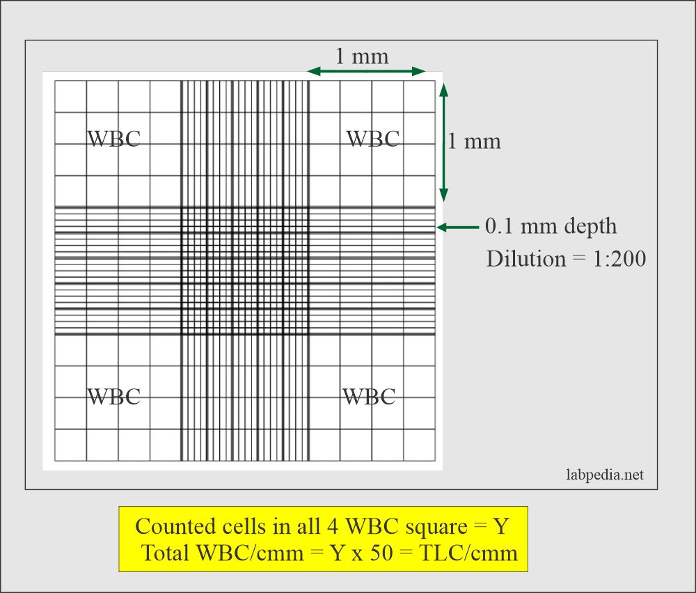
Neubauer chamber counting of white blood cells (TLC)

Procedure to fill the Neubauer chamber

WBCs counted in one of the large squares as a sample
Total leucocyte count Calculations:
- Count the cells in the Neubauer chamber. These are counted in the four large corner squares labeled as WBC and if the number is Y.
- One large area is 1 x 1 mm, and the depth is 0.1 mm.
- Total area counted in 4 large squares = 4 x 1 x o.1 = 0.4 µL (4/10).
- Y x 10/4 is the total WBC in the cell in 1 µL.
- Now dilution is 1:20.
- Number of WBC in 1µL = Y x 10 x 20/4 = Y x 50 = Total WBC count.
- Total TLC = counted cells (Y) x 50 = TLC/cmm.
- If the count is low <4000/cmm, then use the dilution of 1:10.
- The Source of errors are:
- If there are microclots in the sample.
- If inadequate mixing is done.
- Improper filling of the chamber.
- If the dilutions are improper.
- Mistakes in the calculations.
Normal values of total leucocytes are:
Source 2
- Adult /child = 5000 to 10,000 /cmm
- Child ≤2 years = 6200 to 17000 /cmm.
- Newborn = 9000 to 30,000 /cmm
Other sources
- At birth = 10,000 to 25,000/cmm
- Infants = 8000 to 15,000/cmm
- Adults = 4000 to 10,000/cmm
- Pregnant ladies = 12,000 to 15,000/cmm
Increased TLC (Leucocytosis) is seen in:
- Mostly in the case of infections that may be bacterial or viral.
- Localized infections are:
- Meningitis.
- Pneumonia.
- Abscess.
- Tonsillitis.
- Generalized infections:
- Septicemia.
- Acute rheumatic fever.
- Cholera.
- Localized infections are:
- In the case of leukemias.
- After the strenuous exercise.
- Pain and anorexia.
- Epileptic seizures.
- Emotional reaction.
- Mild leucocytosis in pregnancy.
- Acute hemorrhage.
- Intoxications like:
- Poisoning by drugs, chemicals, and venoms (black widow spider).
- Metabolic diseases uremia, acidosis, eclampsia, and acute gout.
- Parenteral proteins and vaccines.
- Acute hemolysis of red blood cells.
- Myeloproliferative diseases.
- Tissue necrosis:
- Burns.
- Gangrene.
- Necrosis of the tumor.
- Acute myocardial infarction.
- Necrosis due to bacteria.
- Physiologic conditions are:
- Emotional stress.
- Excercise.
- Obstetrical labor.
- Menstruation.
Decreased leucocytosis (neutropenia) is seen in:
- This may be seen in fever, malaise, and chills.
- Bacterial Infections.
- Bacterial.
- Septicemia.
- Miliary tuberculosis.
- Typhoid fever.
- Paratyphoid fever.
- Tularemia.
- Brucellosis.
- Viral infections are:
- Hepatitis.
- Influenza.
- Infectious mononucleosis.
- Psittacosis.
- Rubella.
- Measles.
- Hematological diseases:
- Aleukemic leukemia.
- Pernicious anemia.
- Gaucher’s disease.
- Felty’s syndrome.
- Aplastic anemia.
- Deficiency of vitamin B12.
- Drugs and chemicals:
- Antibiotics.
- Analgesics.
- Sulphonamides.
- Antithyroid drugs.
- Arsenicals.
- Marrow depressant.
- Malignant infiltration of the bone marrow.
- Bone marrow aplasia.
- Bone marrow depression by radiations.
- Autoimmune diseases like SLE.
- Critical value = <2500 or >30,000 /cmm ( These are panic value).
- Please see for more details in complete blood count (CBC) part 1.
Very essential information
Thanks.
Sir plz upload lecture on leukemia
I am working on acute leukemias. You will find daily updates.
I have updated acute leukemias. I hope you will like it.
Send a WBC calculation process
Please see at the end of “White blood cell. part 2”.
You made this topic very easy for me
Thanks.
It is so helpful for me
Thanks.
It is 100% easy than to read it from university manual.
Thanks.
Perfectly OK sir
Thanks.
What is 10 in wbc count formula?
Total area counted in 4 large squares = 4 x 1 x o.1 = 0.4 µL (4/10).
x10 comes from above formula.
How about when the number are very high like erythrocytes we use the R squares.
When you have a chance, Can you please elaborate on that? We use it to count sperm concentration of frozen samples used for artificial insemination.we dilute the sample 1:200.
Normal concentrations can range between 100.000 to 2’000.000 per cc.
Please see this link, I hope this may help you.
https://www.labpedia.net/semen-part-1-semen-analysis-semen-examination-and-semen-counting-procedure/
That was exactly what i was looking for. Thank you !
We were correct but wanted to double check. use a dilution 1:200 so will add one extra 0.
Frozen semen is stored under liquid nitrogen that is -196C or 320F and will deteriorate at temperatures over -140C or -220F so that referral page of frozen semen you gave me the frozen semen at -20 should be corrected. I hope this can be my 5 cent contribution to your page…. if you want to.
Thanks again. Great work
Thanks, I am happy that the issue is solved.
Please what is the implications of ovefilling the chamber
And what is the implications of trapping air bubbles in the chamber
You will get the wrong result because of the dilution inconsistency.
Same here, I found it very useful
Thank You Sir
Welcome
I’m really confused on how to count the WBC
Please follow the instructions given on this topic.
https://labpedia.net/white-blood-cell-part-2-total-leukocytes-count/
Thank for your effort on
Thanks.
How can we control a typhoid in africa
1. Good hygiene.
2. Mostly, the cooks are carriers; they need to be given antibiotics. Ampicillin is the most successful antibiotic and should be used in large doses.
3. The oral vaccine in 3 doses is the best way to avoid typhoid fever. Everyone at home should use this vaccine dose every year.
Very useful information, thank you.
Thanks.
Is the concentration of WBC obtained from the RBC lysate method and Ficol gradient method from a healthy individual are same? If not what are values.
Please see this reference.
https://jnm.snmjournals.org/content/58/supplement_1/810
what is clinical significance of this test?
Total leukocytes count differentiates acute and chronic infections. TLC helps to reach the diagnosis of various diseases.
Very intresting indeed
Thanks.
Thanks 👍
Thanks.