Alpha-Fetoprotein (AFP), α-Fetoprotein and its Significance
Alpha-Fetoprotein (AFP)
Sample for Alpha-Fetoprotein (AFP)
- This test is done on the serum of the patient.
- Serum may be stored at 2 to 8 °C for 24 hours; otherwise, freeze it.
- How to get good serum:
- Take 3 to 5 ml of blood in the disposable syringe or the vacutainer. Keep the syringe for 15 to 30 minutes at 37 °C and then centrifuge for 2 to 4 minutes to get the clear serum.
- Mother serum may be taken between 13 to 16 weeks of gestation.
- No special preparation is needed.
Indications for Alpha-Fetoprotein (AFP)
- AFP is used as a screening test for increased risk of fetal defects like:
- Neural tube defect.
- Fetal body wall defect.
- Chromosomal abnormalities.
- In the case of neonatal hepatitis, where the level is >40 ng/mL from the neonatal biliary atresia (level is <40 ng/mL).
- It is advised for placental diseases during pregnancy.
- This is also used as a tumor marker for various tumors, especially liver cell carcinoma.
- AFP is the tumor marker for liver cell carcinoma and germ-cell (non-seminoma) carcinoma.
- It is ordered in the cancers of the ovaries and testes.
- This is a good marker for the follow-up and prognosis of liver cell carcinoma and to monitor the therapy.
Definition of Alpha-Fetoprotein (AFP)
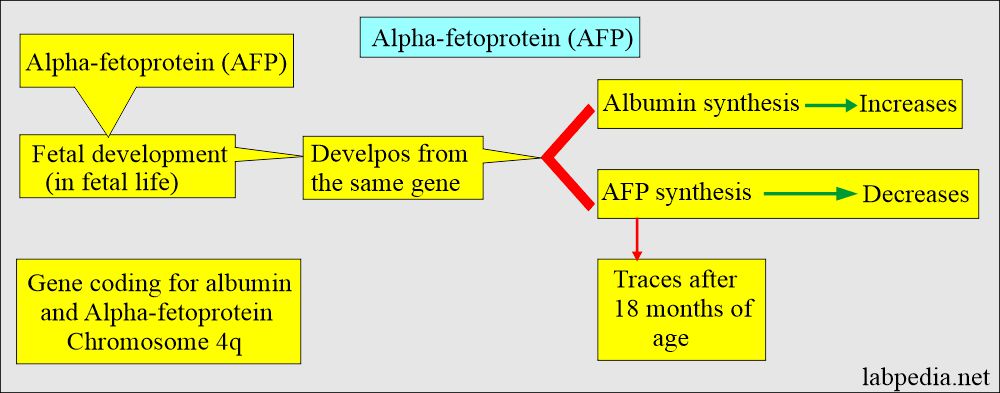
- AFP (α-Fetoprotein) is a principal protein in the fetus, and its function in the adult is unknown.
- AFP is the most significant protein found in the second trimester of the fetus.
- This is a transport protein produced by the fetal liver with functions similar to albumin in infants and adult body fluids.
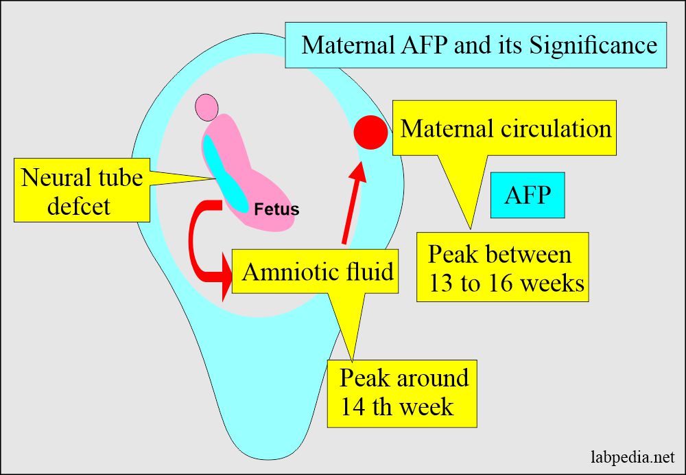
- Due to its large molecular weight, it is found in amniotic fluid and maternal circulation in a minimal amount, so it cannot cross the fetoplacental circulation.
Alpha-Fetoprotein (AFP) structure:
- AFP is a glycoprotein, and it is produced by the fetal liver, gastrointestinal tract, and yolk sac.
- AFP is an oncofetal protein produced by the fetal liver and yolk sac.
- Its molecular mass is 70 kD.
- It consists of a single polypeptide chain and a 4% carbohydrate.
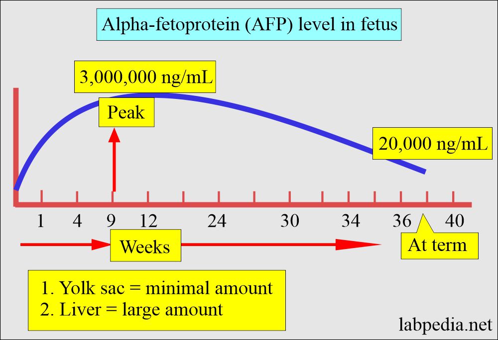
- It is produced in large quantities in fetal life from the fetal yolk sac and liver.
- It is related both genetically and structurally to albumin.
- The gene coding for both is localized to chromosome 4q.
Alpha-Fetoprotein (AFP)level in the fetus:
- The peak production in the fetus is by 13 weeks of gestation, then declines.
- The AFP may be found in the mother’s serum and amniotic fluids.
- AFP level is high in twins and multiple fetuses.
- The Modestly raised level may be seen in the regenerative process of the liver.
- AFP may serve to monitor the course of liver cell carcinoma treatment.
- The AFP level correlates with the tumor size in embryonal malignant teratoblastoma of testes and ovaries, so its measurement is helpful for the monitoring course of the disease.
- AFP is raised in 70% of liver cancer.
Functions of Alpha-Fetoprotein (AFP):
- AFP’s role is to bind and transports substances that are not water-soluble such as:
- Steroids hormones.
- Lipids.
- Vitamins.
- Bilirubin.
- Maternal AFP is lower than the expected value for Down’s syndrome.
- It is a higher level of AFP in neural tube defects.
The normal value of Alpha-Fetoprotein (AFP)
Source 1
| Age | mg/dL |
|
|
|
|
| ng/mL | |
|
|
|
|
|
|
| Maternal | |
| Week of gestation | ng/mL |
|
|
|
|
|
|
|
|
|
|
|
|
|
|
|
|
Source 2
- Adult = <40 ng/mL
- Child < 1 year = <30 ng/mL
Another source
| The level at different ages | Alpha-fetoprotein (AFP) level |
|
|
|
|
|
|
|
|
|
|
|
|
Significance of Alpha-Fetoprotein (AFP):
- It is a marker of Liver cell carcinoma, where greater than 500 ng/dl is found in more than 70 to 80 % of the patient.
- AFP greater than 1000 µg/L suggests cancer (>500 ng/mL is diagnostic of hepatoma).
- It is slightly raised in Cirrhosis.
- Lower levels may be found in patients with large metastasis from Gastric or Colon tumors and patients with acute or chronic hepatitis.
- The presence and persistence of a high level above 500 ng/dl in an adult with liver disease and without any obvious GIT tumors is strongly suggestive of hepatocellular carcinoma.
- AFP may be found in the germ cell tumor.
- AFP is raised in pregnant and neonates.
- It is raised in Choriocarcinoma and Embryonal carcinoma.
- Less commonly raised in Pancreatic carcinoma, Stomach carcinoma, Colon cancer, and lung cancer.
- AFP levels are more significant in twin pregnancies.
- The mother serum raises AFP levels in neural tube defects like spina bifida, anencephaly, myelocele, and hydrocephalus.
- Ultrasonography can confirm this, and the pregnancy may be terminated if the defect is serious.
- AFP level is estimated from 13 to 16 weeks of pregnancy when the fetal production of AFP is highest.
- Adult increased level indicates hepatocellular tumor.
- An increased fetal level indicates a neural tube defect.
Neural tube defects and alpha-fetoprotein (AFP)
- Neural tube defects occur in the early first trimester as the spinal cord and brain develop from an embryonic structure called the neural tube.
- Failure of the neural tube to fuse leads to:
- Spina bifida is also known as meningomyelocele. There is a congenital opening in the spinal cord membrane through which the cord protrudes.
- Encephalocele is a central nervous system defect involving the brain. There is a congenital opening in the skull with protrusion of the brain tissue.
- Anencephaly occurs when the fetus does not develop the cerebrum. There is a greatly reduced brain, particularly the cerebrum, resulting from the failure of the neural tube to close during organ formation.
- Amniotic AFP is more accurate in diagnosing neural tube defects in early gestation ( around 14 weeks) than maternal serum AFP.
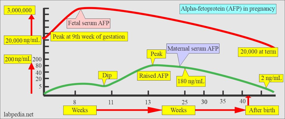
- The AFP at the 8th week is very high; then there is a dip at 11 weeks and again peaks at 13 weeks. Then fall in a long-linear fashion until 25 weeks.
Diagnosis of neural tube defect:
- Before 14 weeks, AFP helps to diagnose neural tube defects. AFP is more than double the normal value.
- High-resolution ultrasound can find this defect.
- There is an increased level of AFP in the amniotic fluid.
- Normal AFP <2.5 Multiples of the median (MoM).
- For the neural tube, the defect is >2.5 MoM.
Down’s syndrome and alpha-fetoprotein (AFP)
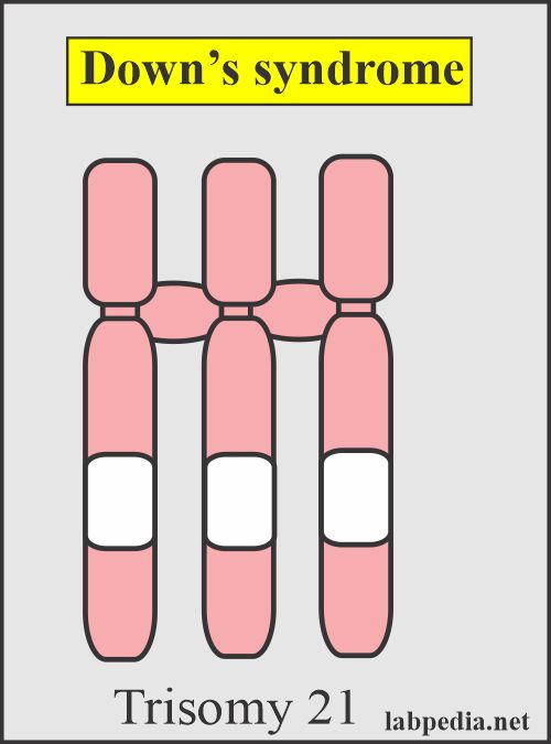
- It is the most common autosomal trisomy.
- Karyotyping shows 47 chromosomes with trisomy in the number 21-22 chromosome group. There are three chromosomes in this group.
- In most patients, caused by translocation., which is seen in chromosome number 14. (called group D)
- It occurs in about 1 in 800 live births.
- It increases with the increasing age of the mother >35 years.
- This is a serious congenital disorder of autosomal chromosome 21 with either trisomy of the long arm or translocation or mosaic of the long arm, mostly in the q21.1 to q22.3 region.
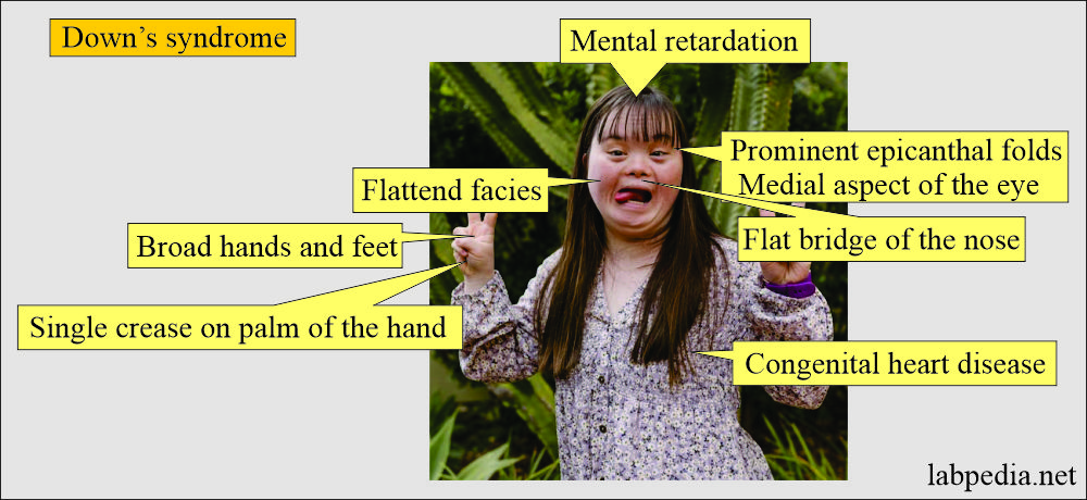
Clinically Down’s syndrome shows:
- Congenital heart defect.
- Muscular weakness.
- Growth and mental retardation.
- Flat facial profile with slanting eyes.
- Broad short skull.
- Broad hands with short fingers.
- An anatomical defect in the esophagus.
Diagnosis of Down’s syndrome(Tripple screening):
- AFP level is low (25% less than the normal level).
- Decreased unconjugated estriol.
- Increased chorionic gonadotropin.
- Quadruple screening when Dimeric inhibin A is added.
- Chorionic villus sampling by the 10th week.
- Karyotyping was obtained from the fetal cells in amniotic fluid by the 18 to 20 weeks of gestation.
Raised alpha-fetoprotein (AFP) has been seen in:
- 100% of the metastatic liver cell carcinoma.
- Screening of liver cell carcinoma. It is raised in 80% of the cases.
- In 50% of nonseminomatous testicular cancer.
- Choriocarcinoma and embryonal carcinoma.
- Noncancerous causes are:
- Cirrhosis.
- Chronic active hepatitis.
- AFP levels are raised in twin pregnancies.
- In 5 to 10% of patients with cirrhosis and hepatitis.
- In alcoholic liver disease.
Decreased Maternal alpha-fetoprotein (AFP) has been seen in:
- Down’s syndrome (Trisomy 21).
- Fetal wastage.
Alpha-fetoprotein (AFP) to diagnose liver cancer:
- AFP is raised for 18 months before the symptoms appear.
- The sensitivity of the AFP for liver cancer is 60%.
- >1000 ng/ml indicates liver cell carcinoma in 50% of the cases, and the tumor size is around 3 cms/.
- 90% of the hepatoma has >200 ng/mL of AFP.
- 70% of the cases have>400 ng/mL.
- A very high level at the diagnosis indicates a poor prognosis.
- If the AFP level does not reach normal after the surgery, it indicates incomplete surgery.
- The postoperative decrease indicates surgical removal, but if AFP again starts rising, it is due to the recurrence of the tumor.
- While 40% of liver cancer may have a normal value, the normal level of AFP does not rule out the possibility of liver cancer.
- Elevated AFP levels may be seen in other conditions as well.
- Although the FDA does not recommend AFP for cancer screening, it detects and monitors liver cell carcinoma and other tumors like testes and ovaries, retroperitoneum, and mediastinum.
Alpha-fetoprotein (AFP) as a tumor marker for:
- Liver cell carcinoma.
- Testicular germ cell tumors.
- Gastric carcinoma is seen in 18% of the cases.
- Pancreatic carcinoma is seen in 123% of the cases.
- Bronchogenic carcinoma is seen in 7% of the cases.
- Colon carcinoma is seen in 7% of the cases.
- Other possible causes of raised AFP:
- Nonseminomatous germ cell tumor.
- Testicular Teratocarcinoma is seen in 75% of the cases.
- Yolk sac tumors.
- Germ cell tumors of the ovaries and testes:
- Embryonal cell carcinoma (value increased in 27% of the cases).
- Malignant teratoma (value increased in 60% of the cases).
- Sometimes may be seen in:
- Hodgkin’s lymphoma.
- Lymphoma.
- Renal cell carcinoma.
Questions and answers:
Question 1: What is the significant level for liver cell carcinoma?
Question 2: What is the recommendation for AFP by the FDA.

