Malarial parasite:- Part 3 – Plasmodium vivax, Benign Tertian Malaria
Plasmodium vivax
Sample for Plasmodium vivax
- Malarial parasites (MP) may be diagnosed with a fever from a patient’s blood smear.
- The best time to make a smear is during shivering.
- Make thick and thin blood smears.
- Serum needed for a Serological method and for PCR.
Definition of Plasmodium vivax
- There is a fever every 48 hours, and it infects young RBCs.
- There may be a relapse for five years.
Why is it called Benign tertiary malaria due to Plasmodium vivax?
- Plasmodium vivax usually causes an acute self-limiting febrile illness, and there is a spike in the fever every third day.
- There is no complication of death in this case, so-called benign tertiary malaria.
Plasmodium vivax facts
- This is the most predominant parasite in most areas of the world.
- 70% to 90% of cases in most of Asia and S. America.
- 50-60% of cases in SE Asia and the western Pacific.
- 1-10% in Africa.
- Plasmodium vivax is the second most significant species and is prevalent in Southeast Asia and Latin America.
- Plasmodium vivax has the complication of a dormant stage in the liver.
- Plasmodium vivax can become active in the liver without mosquito biting and lead to clinical symptoms.
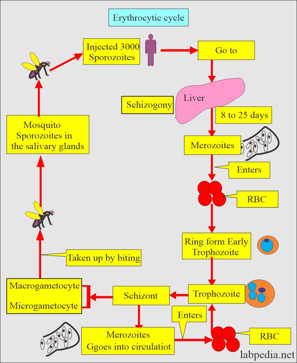
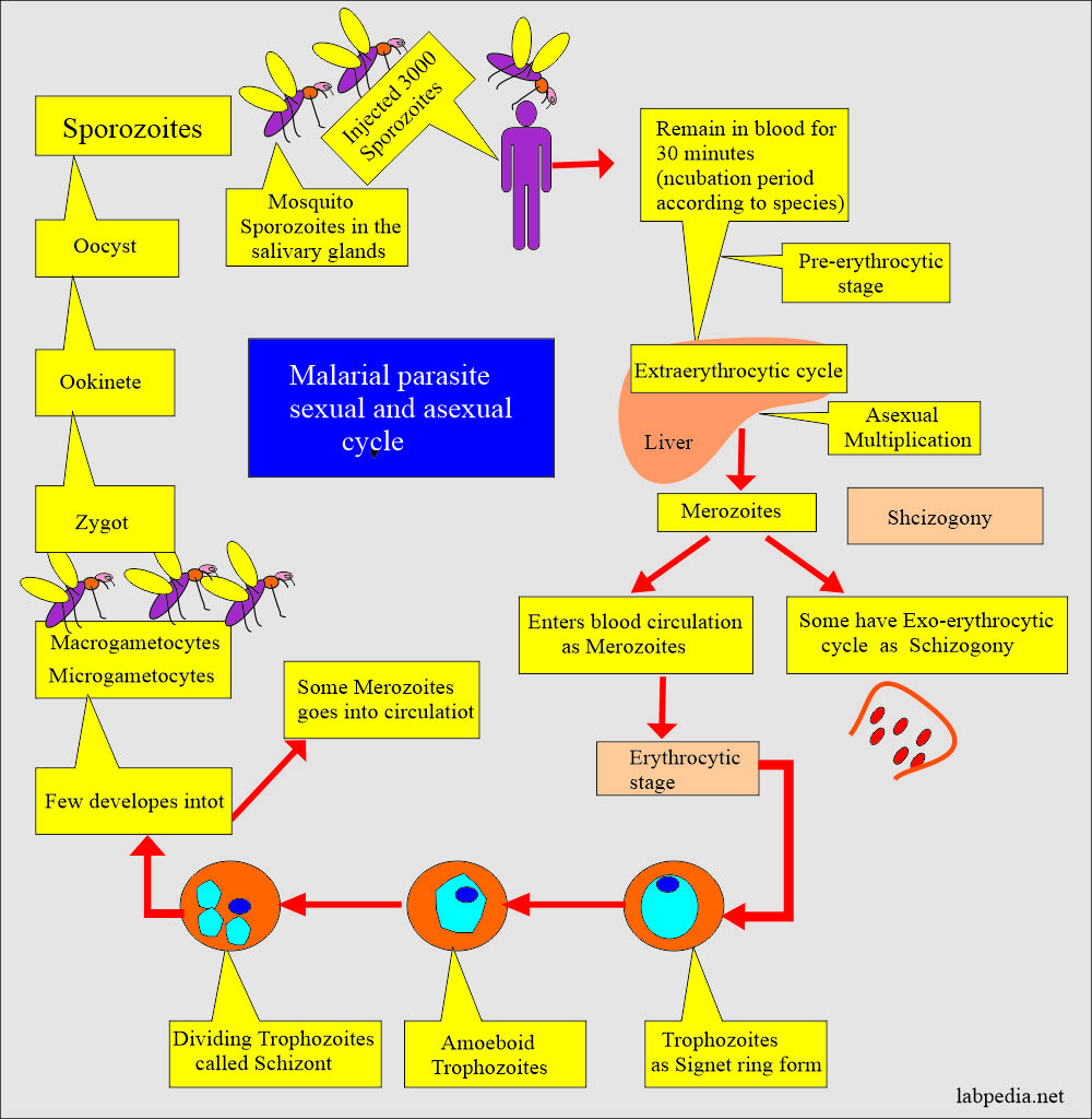
The erythrocytic cycle of Plasmodium vivax
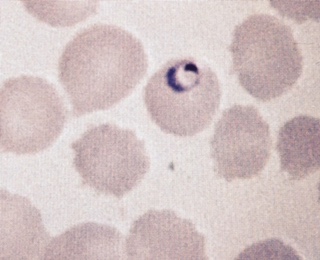
- Plasmodium vivax show ring form with chromatin dot and cytoplasm.
- It measures 1/3 of the RBC.
- Trophozoite may show remnants of the cytoplasm.
- It is an irregular and amoeboid appearance.
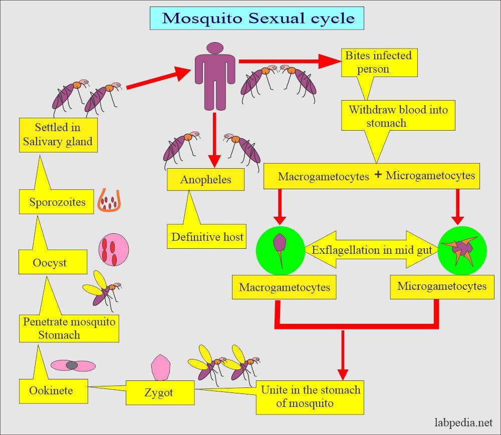
Sexual cycle of plasmodium vivax
- Schizonts are characterized by the presence of multiple chromatin dots.
- Cytoplasm often contains brown pigments.
- There are, on average, 12 to 24 merozoites, with an average of 16.
- These merozoites occupy the majority of RBC.
- Brown pigments may be found.
- Microgametocytes consist of large pink to purple chromatin mass.
- Brown pigments are usually seen.
- Macrogametocytes are characterized by the round to oval homogenous cytoplasm and eccentric chromatin mass.
- A diffuse light brown pigment may be visible throughout the parasite.
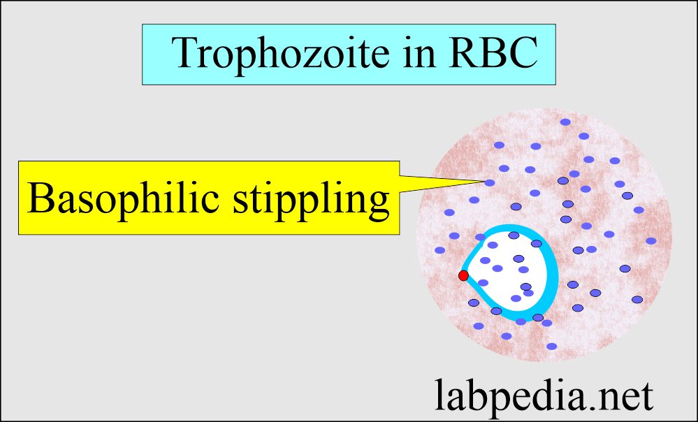
- Schuffner dots are seen with Giemsa stain.
- Infected RBCs are enlarged and distorted.
Clinical presentation of Plasmodium vivax
- The patient develops signs and symptoms after 10 to 17 days of incubation following the infection (bite from the mosquito).
- Early there are flu-like symptoms.
- There may be nausea, vomiting, headache, muscle pain, and photophobia.
- The toxins of the parasite give rise to paroxysm, which occurs every 48 hours.
- If the infection becomes chronic, then it may cause damage to the kidney, brain, and liver.
Diagnosis of Plasmodium vivax
- History of the patient in suspected areas.
- Blood smear:
- Make a blood smear when the patient has a fever. Thin and Thick smears are made.
- The thick smear is more helpful in finding M.Parasites.
- The thin smear is good for identifying the type of malarial parasite.
- Collect blood 6 to 8 hourly till 48 hours to declare negative for malaria.
- Giemsa stain is the best choice.
- Serologic methods are based on immunochromatic techniques. Tests most often use a dipstick or cassette format and provide results in 2-15 minutes.
- Polymerase chain reaction (PCR): Parasite nucleic acids are detected using the PCR technique. This is more sensitive than smear microscopy. This is of limited value for diagnosing acutely ill patients because of the time needed for this procedure.
Mosquito control
- Try to eliminate breeding places:
- Fill the vacant land and pump out the water.
- Remove the junk and water-retaining debris.
- Destroy the larvae:
- Clean the drains.
- Try to remove algae from the ponds.
- Add larva-eating fish to the ponds.
- Use of the insecticide:
- The best example is DDT.
- Use of mosquito repellent:
- Pyrethroid repellent.
- N, N- diethyl meta tolbutamide.
- Use of mosquito nets.
- Use clothes to prevent mosquito bites.
- Train people for malaria prevalence.
- Train the people for the detection of malaria, treatment, and follow-up.
Questions and answers:
Question 1: What is the infective stage of the malarial parasite?

