Diabetes Mellitus:- Part 5 – Glucose in Urine (Glucosuria), Glycosuria, Benedict’s solution
Glucose in Urine (Glucosuria)
What sample of Glucose in Urine (Glucosuria) is needed?
- The test sample is urine.
- The best sample after 2 to 3 hours of the meal.
What are the Indications for Glucose in Urine (Glucosuria)?
- To diagnose diabetes mellitus.
- To monitor diabetes mellitus.
- To evaluate the effectiveness of the therapy.
- To diagnose gestational diabetes.
- It is part of a routine urine examination.
What are the precautions for Glucose in Urine (Glucosuria)?
- Can see false-positive tests when any substance present in the urine can reduce the copper in the clinitest strips.
- Can see the false positive tests in the presence of other sugars like galactose, fructose, and lactose.
- Drugs giving false positive tests are acetylsalicylic acid, Ascorbic acid, cephalothin, chloral hydrate, streptomycin, sulphonamides, and aminosalicylic acid.
- Contamination from the oxidizing agents or bleach.
- Improper storage of the urine strips.
- Drugs giving false-negative results are levodopa, phenazopyridine, and ascorbic acid with clinitest strips.
- Multistix detects ≥50 mg/dL; if the amount is less, it will be negative.
- Sodium fluoride causes enzyme inhibition.
- Refrigerated specimens give decreased enzyme activity.
- Some drugs increase glucose in the urine, like diuretics (thiazide), estrogens, isoniazid, lithium, nalidixic acid, nicotinic acid, chloramphenicol, chloral hydrate, cephalosporin, and aminosalicylic acid.
How will you define Glucose in Urine (Glucosuria)?
- Glycosuria is the presence of reducing substances (glucose, galactose, lactose, and fructose) in the urine.
- Glucosuria is the presence of glucose in the urine. This is specific to the diagnosis of diabetes mellitus. Glucosuria may be with hyperglycemia and, in some cases, maybe without hyperglycemia.
Pathophysiology of Glucose in Urine (Glucosuria):
- Examination of urine for glucose is rapid, noninvasive, and inexpensive for screening urine.
- A large number of urine samples can be tested.
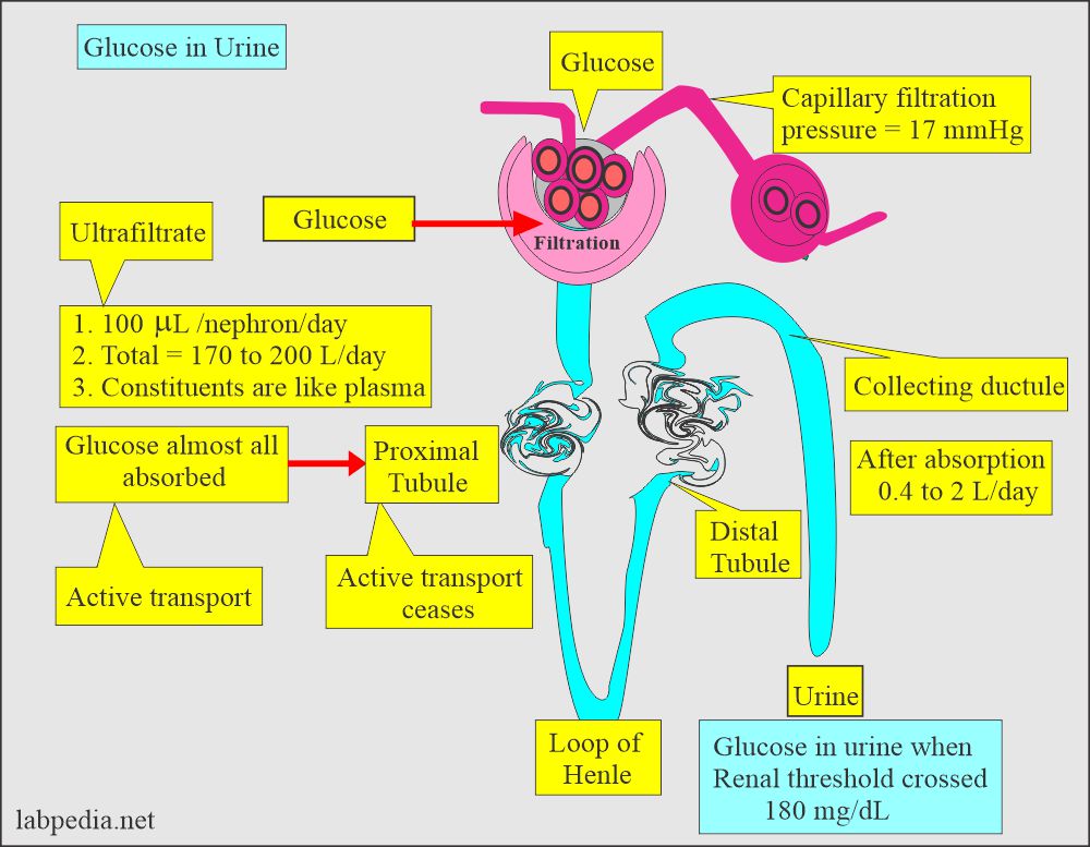
- The glomeruli filter glucose and all of the glucose is reabsorbed by proximal convoluted tubules.
- When glucose exceeds the renal threshold level(>180 mg/dL) of tubules, glucose appears in the urine, called glucosuria. https://labpedia.net/gestational-diabetes-mellitus-oral-gtt/.
- Tubular absorption is an active process to maintain the body’s glucose level.
- Renal threshold for glucose = 160 to 180 mg/dL.
- After the renal threshold values, glucose appears in the urine.
- This is not sensitive nor specific for the control of diabetes because we don’t know about the glucose level below 180 mg/dL.
Name the reducing substances in the urine.
- Lactose.
- Fructose.
- Galactose.
- Maltose.
- Arabinose.
- Xylose.
- Ribose.
What Other reducing substances are found in the urine?
- Uric acid.
- Creatinine.
- Cysteine.
- Ketone bodies.
- Oxalic acid.
- Glucuronic acid.
- Hippuric acid.
- Homogentisic acid.
- Drugs are:
- Ascorbic acid.
- Isoniazid.
- Salicylates.
- Formaldehyde.
What is the normal value of glucose in the urine?
Source 2
- Normally, sugar (glucose ) is absent in the urine.
- Random specimen = negative
- 24 hours specimen = < 0.5 g/day (<2.78 mmol/day).
- Glucose appears in the urine when a blood glucose level of 180 mg/dL or more (crosses the renal threshold).
- Its concentration in the urine correlates with the blood glucose level.
What are the Procedures for reducing substances in the urine?
Benedict’s solution method:
What is the principle of Benedict’s reaction?
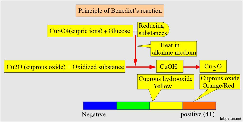
- It is a reaction that is a qualitative method and is very common.
- It contains cupric ions complex to citrate in an alkaline medium.
- Reducing substances convert cupric to cuprous ions.
- It forms yellow cuprous hydroxide or red cuprous oxide.
Benedict’s reagents:
- CuSO4 (cupric sulphate) = 17.3 grams
- Sodium citrate (Na3C6H5O7-2H2O) = 173 grams
- Sodium carbonate (Na2CO3) = 100 grams
- Distle water = 1000 mL
Procedure to prepare the benedict solution:
- Dissolve cupric sulfate in hot water of 100 mL.
- Now dissolve sodium citrate and sodium carbonate with heating water of 800 mL separately. Let it cool.
- Mix solutions 1 and 2 and make up to 1000 ml of water.
- Benedict’s reagent is ready and stable.
Procedure for Benedict’s solution method:
- This can be done on the solution of benedicts as well.
- Take 2.5 mL of Benedict’s solution.
- Add 0.2 ml of urine.
- Place the tubes in a heat block or heat them directly to bring them to 100 °C.
- Examine each tube’s color and the precipitate.
- Different colors develop according to the quantity of sugar in the urine.
- Greenish brown when there is a large quantity >2 g/dL.
Another way to do Benedict’s reaction:
- Benedict’s solution is 5 mL.
- Urine 0.4 mL (8 drops).
- Mix and keep in a boiling water bath for 3 minutes.
Tablet method for Benedict’s reaction:
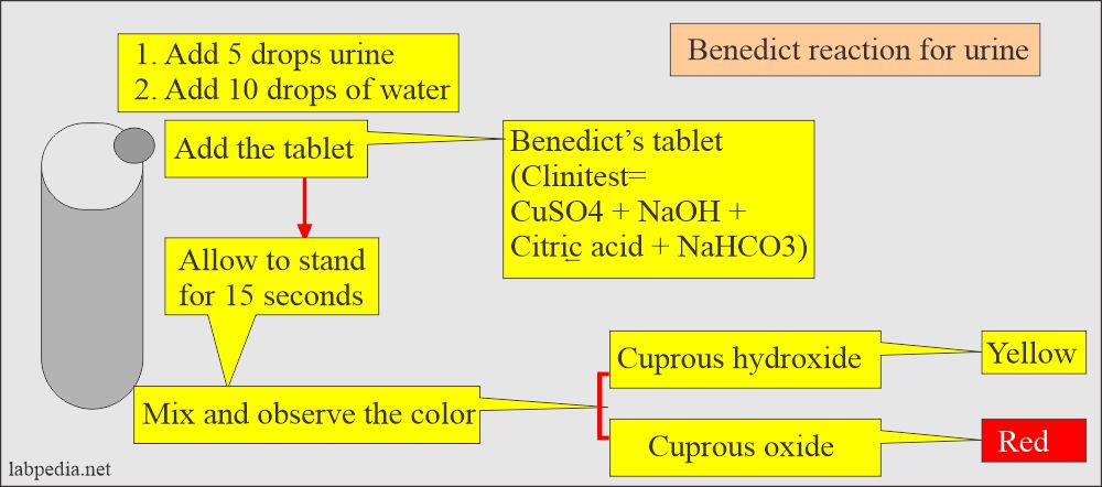
- 5 drops of urine are mixed with 10 drops of water.
- Then add the tablet as known clinitest.
- The procedure for Benedict’s reaction with the tablet (Clinitest) is shown in the following diagram.
- Also, run the negative control as well.
Reporting the result:
- It can be reported as plus + signs, from 1+ to 4+.
- It can report a percentage of 1% to 2 %. This reporting is more accurate.
| Color of the urine after Benedict’s reaction | Reporting method |
The concentration of glucose |
| Blue, clear, or cloudy (Benedict’s solution color) | 0 | NIL |
| Green and no precipitate (may see precipitate) | 1+ | Traces |
| Brown and cloudy | 2+ | Around 0.5 g% |
| Orange and cloudy | 3+ | Approximately 1. o g% |
| Red and cloudy | 4+ | About 2.0 g% or more |
A semiquantitative method for glucosuria:
- This method with different strips, like Clinistix, Diastix, and Chemstrip, is available.
- In all the above strips, the glucose-specific enzyme glucose oxidase is used.
- This is more specific for glucose than Benedict’s method.
- This test is positive when glucose concentration is 100 mg/dL or more.
What is the sensitivity of the commercially available Discs?
- Multistix = 75 to 125 mg/dL
- Diastix = 75 to 125 mg/dL
- Chemstrip = 40 mg/dL in 90% of the specimen.
What are the drawbacks of the strips?
- Urine strips detect mainly glucose, so there are chances for false-negative results due to interfering chemicals in the urine.
- False-positive results are seen in the following:
- If the detergents contaminate the container.
- In very dilute urine, traces may be seen due to sensitivity at low specific gravity.
- If the strips are exposed to air due to improper storage.
- False-negative results are seen in the following:
- It is seen in the intake of vitamin C (ascorbic acid) and tetracyclines.
- In case there are high ketone bodies (≥40 mg/dL) and low glucose levels (75 to 125 mg/dL).
- Sodium fluoride inhibits the enzyme reaction, so it should not be used in the urine as a preservative.
- If urine is refrigerated, it will give false-negative results because of decreased enzyme activity, bringing the urine to room temperature.
Describe the quantitative method for Glucose in Urine (Glycosuria):
- This method uses hexokinase or glucose dehydrogenase procedures.
What are the causes of Increased glucose in urine?
- Diabetes mellitus.
- Renal glycosuria.
- Hereditary defects in the metabolism of other reducing substances like galactose, pentose, and fructose.
- Pregnancy.
- Liver diseases.
- Pancreatic diseases.
- Thyrotoxicosis.
- Cushing’s syndrome.
- Acromegaly.
- Brain injuries.
- Shock.
- Fanconi’s syndrome (Tubular defect).
- Advanced renal tubular diseases.
- Nephrotoxic chemicals like carbon monoxide, lead, and mercury.
The false-negative result is seen in the following:
- Mostly seen due to drugs.
- Ascorbic acid.
- Levodopa.
- Phenothiazine.
Comparison of Benedict reaction and Oxidase method:
| Characteristics | Benedict reaction (CuSO4) | Glucose oxidase |
| Minimum level detected | Glucose 50 to 250 mg/dL | Glucose 50 mg/dL |
| Other sugars detected |
|
Specific for glucose |
| False-negative |
|
|
| False-positive |
|
Questions and answers:
Question 1: Name false positive result for renal glycosuria?
.
Question 2: What is the difference between Benedict's reaction and glucose oxidase method?
- Please see more details on Fasting Blood Glucose.


